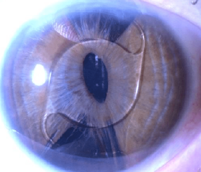
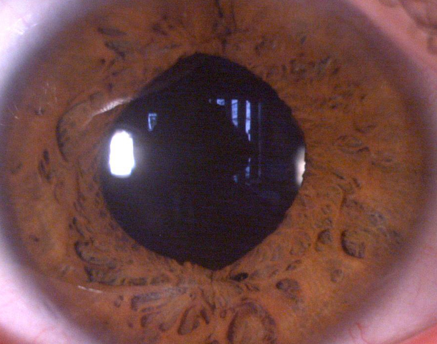
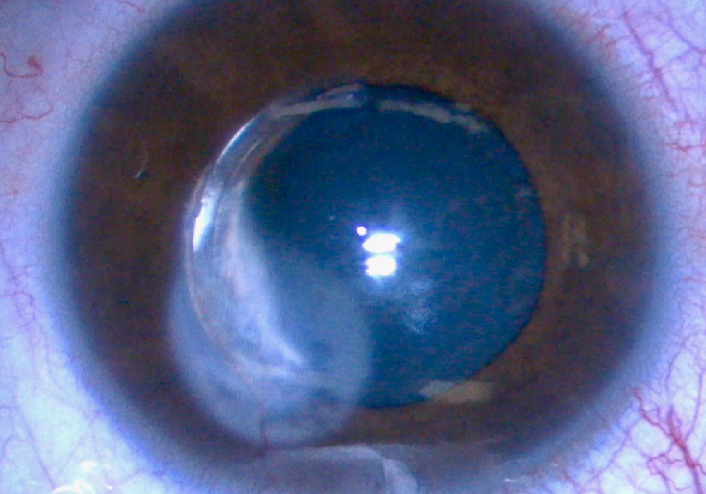
INTRACORNEAL RING REMOVAL 4 YEARS LATER


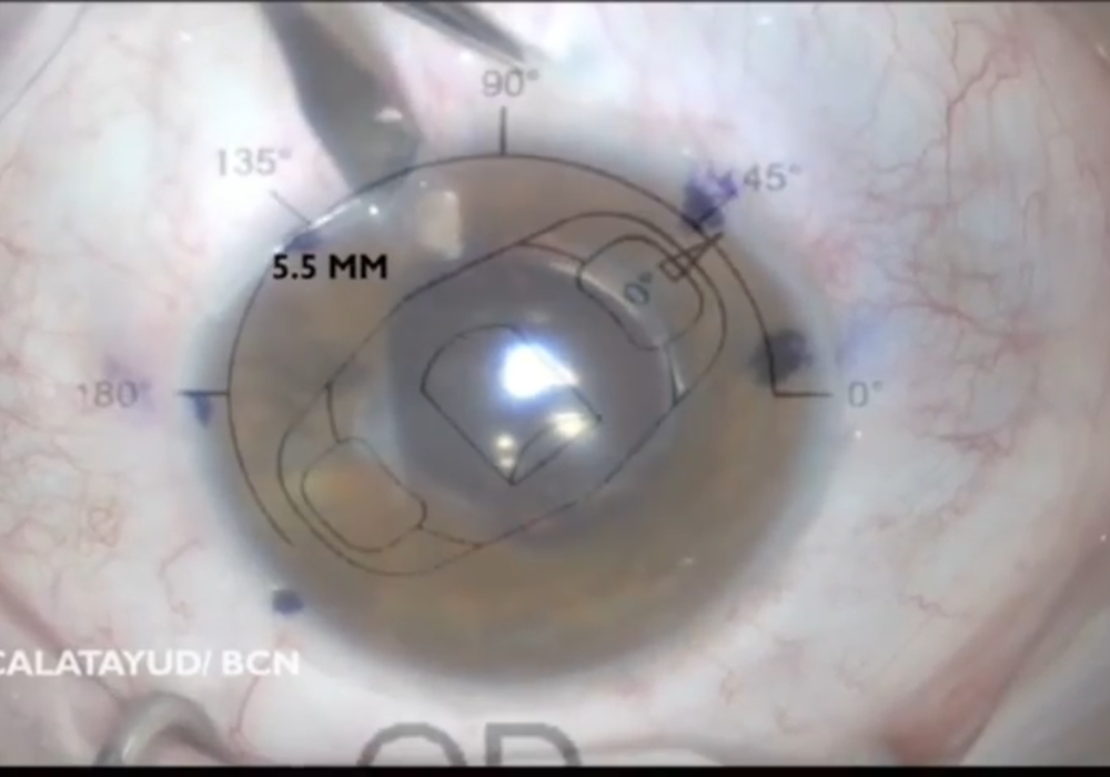
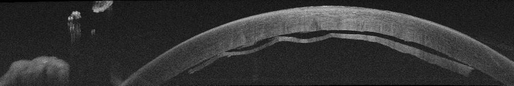
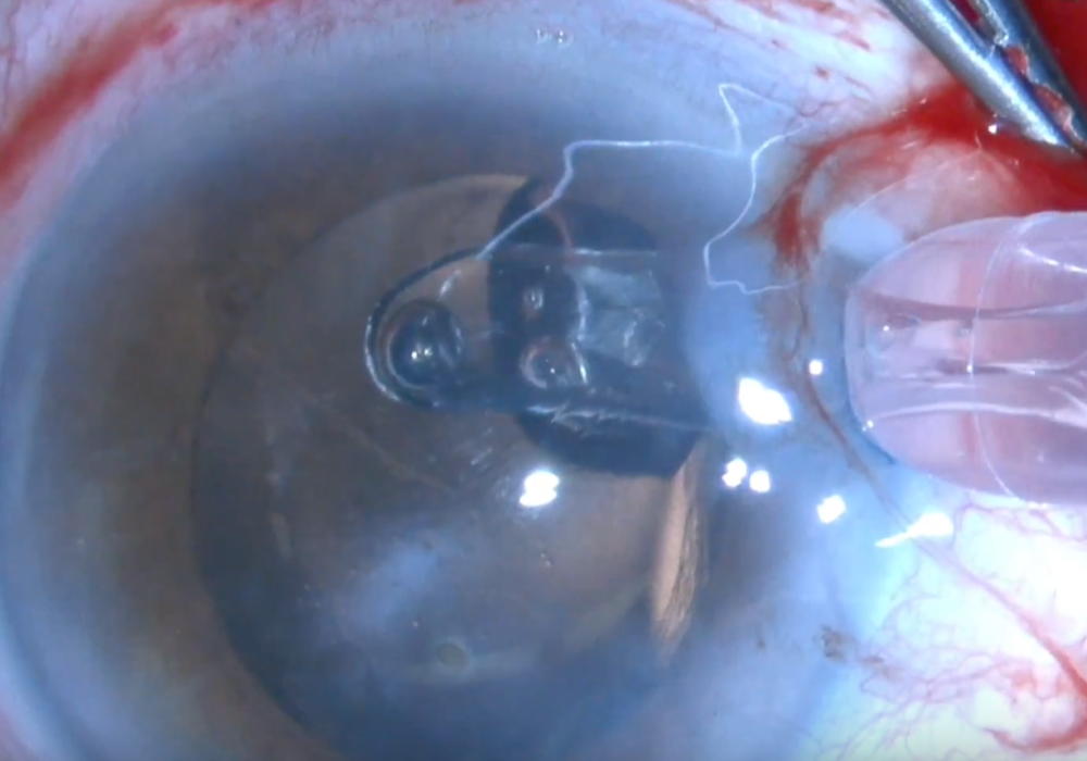
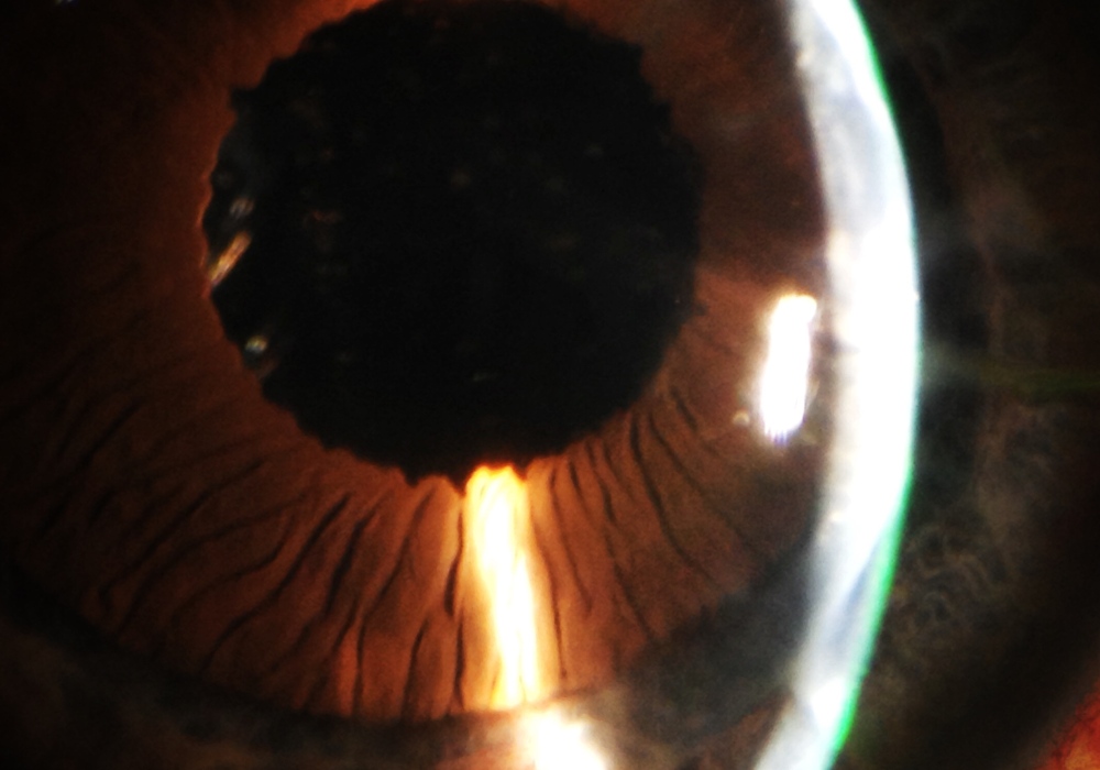
Challenging removal of a fibrotic and vascularised intracorneal segment. Four year after te implantation, the patient reports a white growing spot in the cornea. The Keraring is partially vascularised and covered by a fibrous tissue but not extruded. We tried several maneuvers to remove it, pulling from the hooked hole with an inverted simskey, pushing the distal area, or trying to dissect the primary incision… all of them useless. The only way to do it is looking at the previous records, making a new incision 2 mm from the end of the segment at the depthness set in the initial surgical plan and dissect with a blunt instrument over and under the segment, to refresh the tunnel and pull from one of the sides. No need of specific instruments, buy just in case, fill the tunnel with antibiotic and suture the incisions…

Restoring the eye after severe trauma. Combining Iris prosthesis with aphakia Artisan IOL and vitrectomy to obtain good results

A 14y-o boy with traumatic partial aniridia, aphakia and epiretinal membrane is treated to restore the visual acuity and the anatomy of the globe. There was a previous vitrectomy to remove the intraocular metal foreing body ( a pellet). We start with thep osterior pole, dying the posterior hyaloid with trimacinolone, disecting it and then using dual blue to stain the membrane and peel it off. Once the vitrectomy is finished, we resore the anterior segment with a gently dissection of the iris stroma, trying to preserve as much as possible tissue to be enough to implant an Artisan IOL. A humanoptics artificial iris prosthesis is then cut to 10 mm diameter with a peripheral iridectomy to avoid glaucoma in the postoperative period. We locate the prosthesis into the sulcus, and hook the aphakia IOL under the iris remains. The best corrected visual acuity after one month is 20/20 after the macular edema improves and the stitches are removed. Traumatic eye surgery is an exciting subspecialty which requires requires an experienced surgeon capable of making decisions based on previous knowledge and the correct use of combined intraocular prostheses to obtain the best anatomical and visual results.

Baikoff IOL removal + cataract surgery + pupilloplasty. How to do it without damaging the cornea and the iris

Baikoff angel-supported IOL was implantes during the 90´s to correct high myopia with good visual results but after 20 years we now must confront the fact that all these phakic intraocular lenses must be removed due to late complications like endothelial cell loss, iris-related complications, glaucoma and others like synechiae. Here is one example: a patient with ovalization of the pupil, halos, severe decrease of the endothelium cell population and inicial cataract.

This is one of the reasons why I like ICL from STAAR

iOCt in Lamellar Corneal Transplantation

After a long the working on it, finally there has been released an e-book that I am proud of: iOCT in lamellar corneal transplantation. This is the reason why the blog was on “pause” for some months. I leave you the link to see it.
Hope you enjoy it!!!
https://itunes.apple.com/es/book/oct-intraoperatoria-en-cirug%C3%ADa-lamelar-corneal/id1282060997?mt=11

http://kekulebooks.com/tienda/oct-intraoperatoria-en-cirugia-lamelar-corneal/

And soon in english!!!!

ARTISAN TORIC IOL IN AN INTRAOCULAR LENS EXCHANGE

This is a patient who suffered an intraocular lens rotation after a capsulotomy. The axis turn 90 degrees , so we had to remove the toric IOL an insert another one. Because of the capsulotomy we practice an anterior vitrectomy usin triamcinolone to identify any vitreous in the anterior chamber.

Artisan customized toric IOL is implanted following a specific surgical plan, as described in the video

Transfixing suture in DSAEK

We present a case of a patient with Fuchs’ corneal posterior dystrophy associated to a map-dot-fingerprint anterior dystrophy and a previously failed DMEK , which was treated with DSAEK . After 1 week , the graft began to peel off in an area that had previously suffered a traumatic abrasion of the predescemetic Dua´s layer . We decided to perform a transfixing suture asociated to rebubbleing to ensure the proper adhesion of the graft , since the latter was the third surgery in this patient . Tips and videos that can be seen explicitly showing the technique and the postoperative OCT. Presentamos un caso de una paciente con distrofia corneal posterior de Fuchs asociada a una distrofia anterior tipo mapa-punto-huella, y una DMEK fallida previa, en la que realizamos una DSAEK. Después de 1 semana, el injerto empezó a despegarse en una zona en la que previamente se había producido una abrasión traumática de la capa predescemética de Dua. Decidimos realizar un rebubbleing reforzado con puntos transfixiantes para asegurar la correcta adhesión del injerto, dado que ésta última era la tercera intervención quirúrgica a la que sometíamos a la paciente. Se muestran trucos y vídeos en los que se observa de forma explicita la técnica, así como el OCT postoperatorio.

SMCIS modified for hard cataracts. Tips

Small Incision Cataract Surgery (SMICS) is a very useful method to manage mature cataracts that could be a nightmare with Faco. This case is an only one eye with corneal leucoma an 4+ cataract. We add a limbal temporla incision to mantain the anteriro chamber tight and free of severe changes in the intraocular pressure but use the wide scleral tunnel to remove the cataract protecting the endothelium. We also perform a posterior capsulotomy during the surgery to ensure a good and quick visual rehabilitation and insert the IOL (Lucia from Zeiss) into the bag with the optic ocupying the capsulotomy and the haptics in the periphery. No vitrectomy is needed. The result is a safe and quick surgery with minimal endothelium damage (of course less that the one secondary to a faco in this eye) and a satisifed patient and surgeon.